
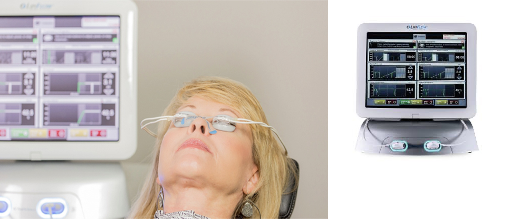
LipiFlow- Meibomian Gland Treatment for Dry Eye
LipiFlow is intended for adults with chronic MGD and evaporative dry eye. LipiFlow is an FDA-approved medical procedure that uses a combination of heat and pressure to treat meibomian gland dysfunction (MGD), a leading cause of evaporative dry eye. This spa like treatment works by unblocking and clearing the glands located in the eyelids that produce the oily layer of the tear film in a simple 12 min in office procedure.
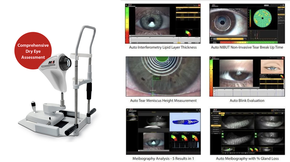
BOSA – Ocular Surface Analyzer
The Bruder Ocular Surface Analyzer (BOSA) is a powerful, advanced diagnostic device designed to evaluate and manage ocular surface diseases (OSD). The diagnostic device has the ability to take a systematic approach to identify, measure, and address these conditions by facilitating targeted and effective management of their ocular health.

RETeval - Electroretinography (ERG)
The RETeval® is a handheld visual electrophysiology device that allows your doctor to detect functional stress in the retina and optic nerve pathways using electroretinography (ERG) and visual evoked potential (VEP) assessments. Think of this like a EKG for your heart, only this is for the retina in the back of your eye. Testing is comfortable, simple, and efficient, using adhesive electrodes to record the electrical impulses from the back of the eye when stimulated by a flash of light. These assessments can help your doctor detect and manage diseases such as diabetic retinopathy, glaucoma, and inherited and acquired retinal dystrophies.

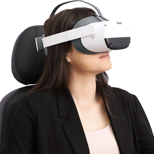
Dark Adaptation - AdaptDx
Dark adaptation is the process of the eyes adjusting to low levels of illumination. During dark adaptation, the sensitivity of the eye to light is increased. The two major components of dark adaptation are dilation of the pupil and photochemical alterations of the retina. Impaired dark adaptation speed indicates retinal cell dysfunction that may lead to macular degeneration.

Optos - Widefield Retinal Imaging
Optos introduced ultra-widefield (UWF™) retinal imaging to enable eyecare professionals to discover, diagnose, document and treat ocular pathology that may first present in the periphery - pathology which may go undetected using traditional examination techniques and equipment. Our UWF, high-resolution retinal imaging devices each image more than 80% or 200˚ of the retina in a single capture

Endothelial Cell Measurement
Our Konan Specular Microscope is the Gold Standard to detect and monitor corneal endothelial cells for the following clinical applications: Glaucoma, Cataract, Refractive Surgery, Corneal Disease, Contact/specialty contact lens fittings.
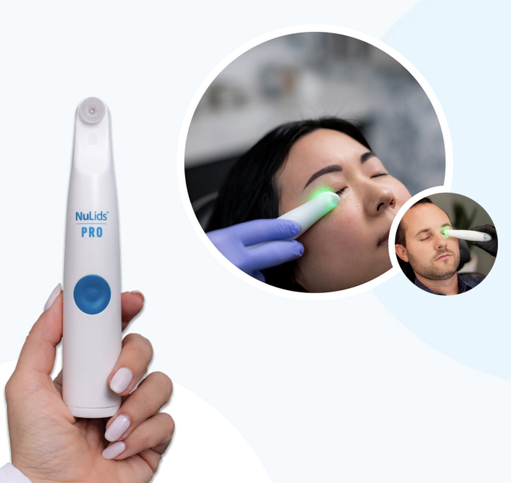
NuLids- Eye Lid Biofilm treatment
NuLids is a FDA Registered, highly effective alternative treatment for dry eye disease specifically designed to gently stimulate and rejuvenate the Meibomian glands that can reduce the signs and symptoms associated with blepharitis and dry eye. A natural way to significantly improve tear stability (and the amount of time it takes for your tears to evaporate) by increasing Meibomian gland output – this means your eyes stay lubricated and symptom-free longer, as nature intended. NuLids is a clinically proven mechanical method to de-cap and stimulate Meibomian glands (like a facial for your eyes- unclogging your glands and stimulating the natural production of oils to keep your eyes lubricated.) NuLids is also very effective in immediate treatment of Demodex blepharitis by directly removing the Demodex mite infestation and cleaning the biofilm layer of the eyelids.
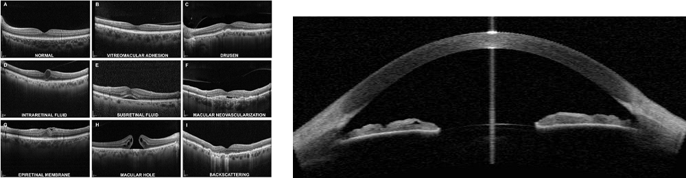
OCT- Retinal and Anterior Segment Analysis
OCT, or Optical Coherence Tomography, allows for fast and precise non-invasive visualization of the retina and optic nerve structure, cornea, iris and the eye’s drainage system. By using light reflection, OCT scans a beam of light to create 3D images that show the retina’s layers, optic nerve and the structures in the front of the eye in microscopic detail.
Detailed OCT images of ocular tissue structure, enabling early detection of anomalies and monitoring of ocular disease progression. OCT is an essential tool for diagnosing and effectively treating ocular health problems.
Visual Fields
Your eyes normally see a wide area of the space in front of you. Without moving your eyes, you can see not only what’s straight ahead, but also some of what’s above, below and off to either side. Providers call all of the area you can see that isn’t right in front of you “peripheral vision.” This surrounds the area that’s right in front of you that you can see (central vision). Visual field testing is an important part of regular eye care for people who are at risk for vision loss from disease and other problems. If your visual field is limited, your ability to drive may be in jeopardy.
A visual field test measures two things:
- visual field test measures your peripheral vision, or how well you can see above, below and to the sides of something you’re looking at. How far up, down, left and right your eye sees without moving your head or eyes. This is generally called Perimetry.
- How sensitive your vision is in different parts of the visual field, which is the name for the entire area that you can see. These Threshold fields tests how much bright the light must be for you to see it.
People with the following conditions should be monitored regularly:
- Glaucoma
- Multiple sclerosis
- Thyroid eye disease (Graves' disease)
- Pituitary gland disorders
- Brain problems (such as a tumor that may be pressing on visual parts of the brain)
- Strokes
- Long-term use of certain medications (such as Plaquenil, or hydroxychloroquine,
- People with diabetes and high blood pressure complications
Our office uses several different devices to measure each of these. , or Optical Coherence Tomography, allows for fast and precise non-invasive visualization of the retina and optic nerve structure, cornea, iris and the eye’s drainage system. By using light reflection, OCT scans a beam of light to create 3D images that show the retina’s layers, optic nerve and the structures in the front of the eye in microscopic detail.
Detailed OCT images of ocular tissue structure, enabling early detection of anomalies and monitoring of ocular disease progression. OCT is an essential tool for diagnosing and effectively treating ocular health problems.
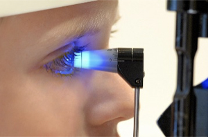
Tonometry
Tonometry is a medical procedure used to measure the pressure inside the eye, known as intraocular pressure (IOP). It is an important test for detecting and monitoring glaucoma, a condition that damages the optic nerve due to high IOP. It is a simple, non-invasive procedure that can help prevent vision loss caused by glaucoma. There are several ways to test tonometry.
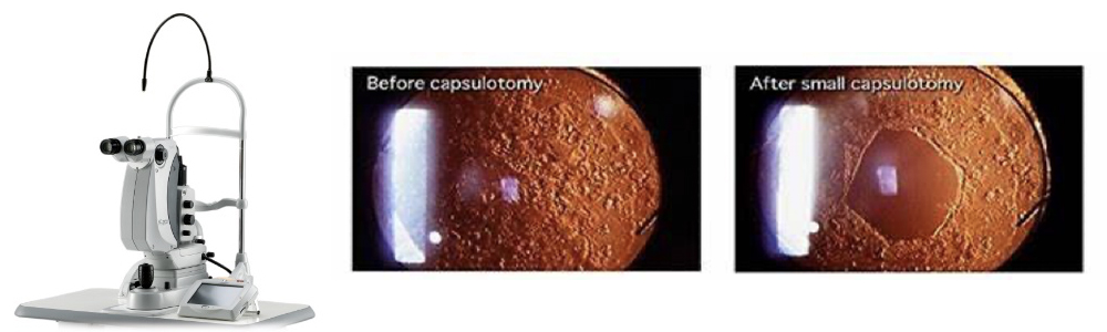
Nidek YC-200 Laser S (Treatment of After-Cataracts & Glaucoma)
A Nidek dual laser refers to an ophthalmic laser system with two distinct laser capabilities, most commonly an Nd:YAG and a Selective Laser Trabeculoplasty (SLT) laser, integrated into a single console. The Nidek YC-200 S plus is such a dual laser, providing both YAG treatment for posterior capsulotomy and SLT for glaucoma, and features high-quality optics and a dual-beam aiming system for precise treatment.
YAG laser capsulotomy is a minimally invasive procedure that uses a laser to create an opening in the posterior capsule of the eye, which is a thin membrane that surrounds the artificial lens (intraocular lens, or IOL) implanted during cataract surgery.
Selective Laser Trabeculoplasty (SLT) is a minimally invasive laser procedure used to treat open-angle glaucoma. SLT is a safe and effective treatment option for open-angle glaucoma. It can provide significant relief from glaucoma symptoms without the need for surgery or long-term use of eye drops.
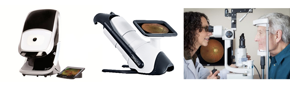
Retinal Photography
Retinal photography uses high-resolution cameras to capture images of the back of the eye, revealing the retina, optic nerve, and blood vessels to detect and monitor eye conditions like diabetic retinopathy, glaucoma, and macular degeneration. The resulting digital images form this non-invasive process provides a permanent, detailed record for early diagnosis, disease progression monitoring, and educating the patient about their eye health. We use several methods to capture these images:
Optos – uses ultra-widefield scanning laser technology to capture a single, high-resolution digital image of the retina, covering up to 82% (200 degrees) in a single shot
Digital Retinography System (DRS) retinal camera is a fully automated, non-mydriatic fundus camera used to take high-quality images of a patient's retina. DRS technology operates without the need for pupil-dilating eye drops (non-mydriatic), a process that saves time and is more comfortable for patients.
Fundus autofluorescence (FAF) is a non-invasive imaging technique used to diagnose and monitor retinal diseases, particularly those affecting the retinal pigment epithelium (RPE). It works by capturing the natural fluorescence, or "autofluorescence," produced by lipofuscin, a metabolic byproduct that accumulates in the RPE cells. The pattern of autofluorescence on the images helps eye care specialists detect and assess retinal health.
Video capture- Each of our Biomicroscopes (Slit Lamps) is attached to a video/ photography system to capture abnormalities for follow up.
Anterior Ocular Photography
A biomicroscope camera provides high-resolution digital imaging of the eye by capturing light from a specialized microscope. The camera can capture images using different lighting techniques, such as retro illumination (light reflecting off the back of the eye) or diffuse illumination (general ambient light), to highlight different parts of the eye.
Slit lamp: The instrument's light source projects a thin, high-intensity sheet of light—the "slit"—into the eye, allowing the doctor to view its internal and external structures in three dimensions.: A camera attached to the slit lamp's eyepiece or an integrated camera captures the image that the doctor sees.
BOSA – this system allows us to capture Blink Rate, tear volume and break up time
VX-120 – this system captures a depth scan of the front of the eye as also any cataracts or after cataracts that could limit vision
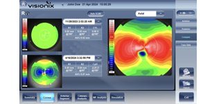
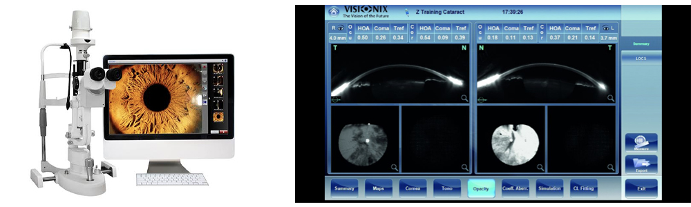
Visionix VX 120 Wave Front Visual Analyzer
This device utilizes wavefront technology to analyze the refractive state of the entire eye by sending a low-power laser into the eye and measuring the shape of the reflected wavefront of light. The device then analyzes the aberrations, or imperfections, in the eye's optical system to create a detailed map. This helps in the early detection of eye pathologies like keratoconus, diabetic retinopathy, glaucoma, and cataracts. The device combines this technology with other diagnostic tools to provide a comprehensive analysis of the patient's visual health.
In addition to refractive (prescription lenses) information the VX-120 also give us insight into the front of the eye, any opacities in the lens, a detailed view of the inside of the anterior portion of the eye and detailed topography of the shape of the eye
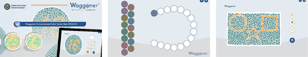
Waggoner Color Vision Test (FAA approved)
The Waggoner color vision tests are a suite of diagnostic and screening tools, including the advanced Waggoner Computerized Color Vision Test (WCCVT), that can detect and categorize both genetic and acquired color deficiencies. They are a modern alternative to traditional tests like the Ishihara plates.
Military and FAA acceptance: The WCCVT is recognized by the U.S. Navy, Army, and Coast Guard as a military-grade test for applicants and personnel. It is also one of only three precision color vision tests approved by the Federal Aviation Administration (FAA).
Pediatric testing: Waggoner's "Color Vision Testing Made Easy" (CVTME) is a component of the suite and uses child-friendly symbols like stars, circles, and squares instead of numbers, allowing for the reliable testing of children as young as three.
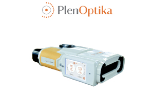
QuickSee Digital Auto Refractor
A handheld autorefractor and keratometer. This portable medical device is used to quickly and accurately measure a patient's refractive errors (such as nearsightedness, farsightedness, and astigmatism. QuickSee allows a binocular and open-view design: Unlike many traditional autorefractors that are closed-view and monocular, QuickSee allows the patient to look through it with both eyes at a distant, real-world target. This helps prevent "instrument myopia," a phenomenon where a patient's eyes focus on the device itself, leading to inaccurate results.
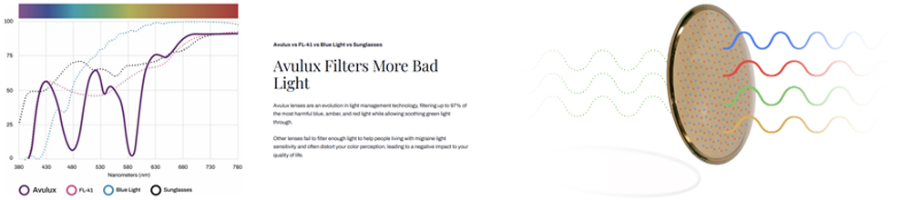
Avulux Migraine & Light Sensitivity Lenses
Avulux migraine glasses are clinically proven lenses designed to filter out specific wavelengths of light that can trigger or worsen migraine symptoms and light sensitivity (photophobia). The lenses appear clear and can be worn indoors and outdoors, and are available in prescription and non-prescription versions.
Avulux lenses are the first to have been tested in a clinical trial and received FDA confirmation of classification to be marketed as general wellness tools, which may, as part of a healthy lifestyle, help people living with migraine. Preliminary research conducted at the University of Utah also indicates that Avulux may help patients with visual snow syndrome and SEES.
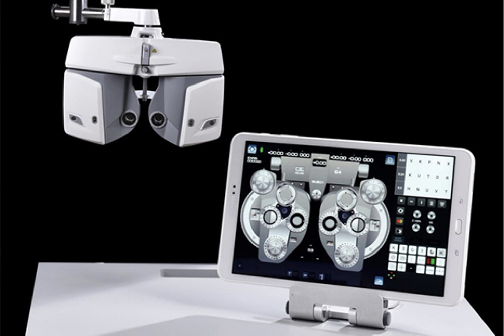
Visionix VX 65 Digital-Computer Assisted Exams
We use a digital exam system (Visionix VX 65) to get the most accurate prescriptions for our patients. This digital system speeds up the exam process for all patients including adults and children. It combines the old standard of a traditional refraction with the speed and flexibility of computer assisted technology.
